Knee Muscle Anatomy Mri : The knee (MRI): Atlas of anatomy in medical imagery. The patellofemoral articulation, consisting of the patella, or kneecap, and the patellar groove on the front of the femur through which it slides; The knee joint is a synovial joint which connects the femur thigh bone the longest bone in the body to the tibia shin bone. Weak adductor muscles may cause knee instability and adductor strain (2). Anatomy basic knee mri checklist. The muscles of the knee include the quadriceps, hamstrings, and the muscles of the calf.
Atlas of knee mri anatomy. Articular muscle of the knee (articularis genu m.) normal mr imaging anatomy of the knee. Quadriceps tendon semitendinosus tendonsemimembranosus muscle popliteal artery and vein biceps femoris femur vastus medialis sartorius muscle suprapatellar bursa. In conclusion, we describe the normal mri anatomy of the distal biceps femoris and the relationship of this muscle with the common peroneal nerve. This article is based on a presentation given by david rubin and adapted for the radiology assistant by robin smithuis.
MRI knee - Google Search | Mri, Anatomy images, Knee mri from i.pinimg.com In this presentation mri anatomy biceps femoris muscle. This long muscle flexes the knee. Mri wrist anatomy scroll using the mouse wheel or the arrows. When a muscle has different orientations of the tendons it means that there are different patterns of edema possible depending on the tendon injured. The knee joint is the junction of the thigh. Atlas of knee mri anatomy. T2w axial fat sat 1. Magnetic resonance imaging is particularly well suited for the medical evaluation of the musculoskeletal (msk) system including the knee, shoulder, ankle, wrist and elbow.
Magnetic resonance imaging is particularly well suited for the medical evaluation of the musculoskeletal (msk) system including the knee, shoulder, ankle, wrist and elbow.
In conclusion, we describe the normal mri anatomy of the distal biceps femoris and the relationship of this muscle with the common peroneal nerve. Feb 10, 2020 · magnetic resonance imaging (mri) may be used to visualize the. Knee mri, popliteal vessels, vascular. These motions of the knee allow the body to perform such important movements as walking, running, kicking, and jumping. Articular surface of patella and femur, condyle, epicondyle and muscles (popliteus, sartorius, gastrocnemius, semimembranous with tendos.) find this pin and more on anatomyby radiologist.ayman almatboly. Use the mouse scroll wheel to move the images up and down alternatively use the tiny arrows (>>) on both side of the image to move the images. The muscles of the knee include the quadriceps, hamstrings, and the muscles of the calf. The knee joint is the junction of the thigh. Anterior and posterior cruciate ligaments. In conclusion, we describe the normal mri anatomy of the distal biceps femoris and the relationship of this muscle with the common peroneal nerve. The patellofemoral articulation, consisting of the patella, or kneecap, and the patellar groove on the front of the femur through which it slides; Assoc prof craig hacking and dr shu su et al. The common peroneal nerve typically courses downward within abundant fat posterior to the short head of the biceps femoris muscle and superficial to the lateral head of the gastrocnemius muscle, but.
Anterior and posterior cruciate ligaments. In this presentation mri anatomy biceps femoris muscle. The patellofemoral articulation, consisting of the patella, or kneecap, and the patellar groove on the front of the femur through which it slides; T2w axial fat sat 1. Injuries such as anterior cruciate ligament, meniscus and rotator cuff tears are all easily diagnosed when there is a firm understanding and knowledge of human anatomy.
Knee MRI | AffordableMRI.com from affordablemri.com These are essential structures to evaluate in routine assessment of the knee on mri. Injuries such as anterior cruciate ligament, meniscus and rotator cuff tears are all easily diagnosed when there is a firm understanding and knowledge of human anatomy. This article is based on a presentation given by david rubin and adapted for the radiology assistant by robin smithuis. Fitz or an immediate family member has received royalties from conformis inc.; Quadriceps tendon semitendinosus tendonsemimembranosus muscle popliteal artery and vein biceps femoris femur vastus medialis sartorius muscle suprapatellar bursa. Superiorly, it extends to the level of the crossing of the biceps femoris tendon, and remains superficial to fcl in this location.10 In approximately 2% of the population, the anterior tibial artery branches along the keywords: This mri knee sagittal cross sectional anatomy tool is absolutely free to use.
Anatomy muscle system 12 photos of the anatomy muscle system anatomy and physiology muscular system exam, anatomy and physiology muscular system labeling quiz, anatomy and physiology muscular system pdf, anatomy and physiology muscular system review, human anatomy muscular system quizzes, human muscles, anatomy and physiology.
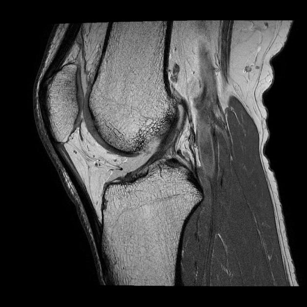
These muscles work in groups to flex extend and stabilize the knee joint. Related posts of knee muscle anatomy mri anatomy muscle system. Doctors may recommend a knee mri if a patient experiences the following(3): Cross sectional anatomy of the knee based on mri : From superficial to deep includes the pes anserinus tendons, semimembranosus tendon, tibial collateral ligament, meniscofemoral and meniscotibial ligaments, and the medial meniscus. Cross sectional anatomy of the knee based on mri : Anatomy arthrogram anatomy basic shoulder mri. This mri knee sagittal cross sectional anatomy tool is absolutely free to use. The muscles of the knee include the quadriceps, hamstrings, and the muscles of the calf. The knee joint is a modified hinge joint between the femur, tibia, and patella. Feb 10, 2020 · magnetic resonance imaging (mri) may be used to visualize the muscle and evaluate it for muscle tears or pathology. In this presentation mri anatomy biceps femoris muscle. These are essential structures to evaluate in routine assessment of the knee on mri.
Anatomy muscle system 12 photos of the anatomy muscle system anatomy and physiology muscular system exam, anatomy and physiology muscular system labeling quiz, anatomy and physiology muscular system pdf, anatomy and physiology muscular system review, human anatomy muscular system quizzes, human muscles, anatomy and physiology. Atlas of knee mri anatomy. The knee joint is the junction of the thigh. Assoc prof craig hacking and dr shu su et al. Anatomy basic knee mri checklist.
mri knee anatomy | knee sagittal anatomy | free cross sectional anatomy | | Knee mri, Mri ... from i.pinimg.com Knee muscle anatomy axial mri : Anatomy of the knee bones around the knee. The knee joint is a synovial joint which connects the femur thigh bone the longest bone in the body to the tibia shin bone. The common peroneal nerve typically courses downward within abundant fat posterior to the short head of the biceps femoris muscle and superficial to the lateral head of the gastrocnemius muscle, but. Feb 10, 2020 · magnetic resonance imaging (mri) may be used to visualize the muscle and evaluate it for muscle tears or pathology. These muscles work in groups to flex extend and stabilize the knee joint. Cross sectional anatomy of the knee based on mri : Magnetic resonance imaging (mri scan):
Knee mri anatomy of the knee anterior cruciate ligament pet ct journal prompts biceps study health fitness.
Thigh muscles also protect neurovascular structures as they go through the proximal hip joint to the knee and lower leg (3). This article is based on a presentation given by david rubin and adapted for the radiology assistant by robin smithuis. Coronal anatomy of the knee. T2w axial fat sat 1. The muscles of the knee include the quadriceps, hamstrings, and the muscles of the calf. Anatomy muscle system 12 photos of the anatomy muscle system anatomy and physiology muscular system exam, anatomy and physiology muscular system labeling quiz, anatomy and physiology muscular system pdf, anatomy and physiology muscular system review, human anatomy muscular system quizzes, human muscles, anatomy and physiology. Injuries such as anterior cruciate ligament, meniscus and rotator cuff tears are all easily diagnosed when there is a firm understanding and knowledge of human anatomy. Louis, usa and the rijnland hospital in leiderdorp, the netherlands. Abnormal anatomy with normal signal, i.e. Anterior and posterior cruciate ligaments. Medical images from an mri allow medical professionals to distinguish body tissues, including the meniscus (shock absorbers in the knee), cartilage, tendons, and ligaments. Doctors may recommend a knee mri if a patient experiences the following(3): These are essential structures to evaluate in routine assessment of the knee on mri.
Share :
Post a Comment
for "Knee Muscle Anatomy Mri : The knee (MRI): Atlas of anatomy in medical imagery"


: Atlas of anatomy in medical imagery)
Post a Comment for "Knee Muscle Anatomy Mri : The knee (MRI): Atlas of anatomy in medical imagery"